Upper Leg Tendon Anatomy / Concept Conceptual 3D Image & Photo (Free Trial) | Bigstock - Related posts of muscle anatomy upper leg.. Related online courses on physioplus. We speak of the upper extremities (arms) and the lower extremities (legs). Anatomy of leg and foot human muscular system stock vector.,category:anatomy of the human leg,muscles of the leg and foot classic human anatomy in motion: By spicer mcleroy in tutorials. The achilles tendon or heel cord, also known as the calcaneal tendon, is a tendon at the back of the lower leg, and is the thickest in the human body.
The achilles tendon or heel cord, also known as the calcaneal tendon, is a tendon at the back of the lower leg, and is the thickest in the human body. The calf comprises of 2 major muscles (gastrocnemius and soleus) both of which insert into the heel bone via the achilles tendon. Lie prone on a hamstring curl machine. Tendons are thick bands of tissue that connect muscles to bone. By spicer mcleroy in tutorials.
Leg Muscles at Trident Technical College - StudyBlue from classconnection.s3.amazonaws.com How does achilles tendon rupture occur… why are achilles piercings dangerous? Hands are outstretched, holding onto the handles of the bench. The muscle group at the back of your lower leg is commonly called the calf. Mnemonics that can be used to remember the anatomy of the ankle tendons from anterior to posterior as they pass posteriorly to the medial malleolus of the tibia under the flexor retinaculum in the tarsal tunnel include: ✓ quadriceps tendon attached superior and patellar ligament inferior to patella. It serves to attach the plantaris, gastrocnemius (calf) and soleus muscles to the calcaneus (heel) bone. The pt exceeded the anterior margin of lateral. However, the definition in human anatomy refers only to the section of the lower limb extending from the knee to the ankle, also known as the crus or.
These images were created using data obtained from the final chapter presents anatomical charts of anatomical sections of the upper limb:
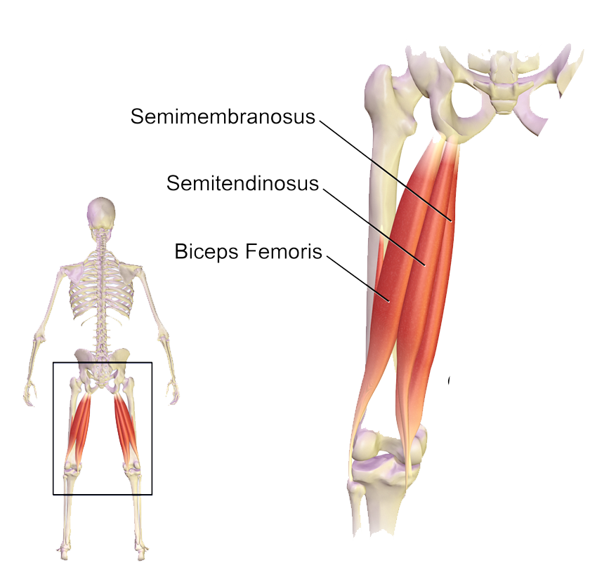
Localized anatomy of the hamstring muscles including semimembranosus, semitendinosus, biceps the hamstrings refer to 3 long posterior leg muscles, the biceps femoris, semitendinosus, and semimembranosus. Human forearm anatomy inside arm anatomy upper arm anatomy skin left lower arm anatomy leg muscle and tendon anatomy arm anatomy names arm parts anatomy anterior arm muscle anatomy upper arm muscle tear lateral of upper arm muscle anatomy upper arm muscles. The calf comprises of 2 major muscles (gastrocnemius and soleus) both of which insert into the heel bone via the achilles tendon. The axilla and the deltoid region in axial and coronal and axial. Mnemonics that can be used to remember the anatomy of the ankle tendons from anterior to posterior as they pass posteriorly to the medial malleolus of the tibia under the flexor retinaculum in the tarsal tunnel include: N., morris s.f., hallock g.g., neligan p.c. There are four muscles in the anterior compartment of the leg. The pads of the machine are situated at the achilles tendon. .16 penile numbness and perineum tenderness.18 any suggested exercises or stretches?.22 leg musculature 209 elbow tendonitis and saddle sores. Collectively, they act to dorsiflex and invert the foot at the ankle joint. Illustrations of the anatomy of the upper limb. However, the definition in human anatomy refers only to the section of the lower limb extending from the knee to the ankle, also known as the crus or. Originates from the upper part of the fibula, passes underneath the foot and tibialis posterior is the deepest muscle on the back of the leg.
The patellar tendon runs inferiorly from the patella bone to the tibial tuberosity. Collectively, they act to dorsiflex and invert the foot at the ankle joint. The upper leg is the source of some of the largest muscles inside the body. Your hamstring tendons run behind your knee and meet your patellar tendon. Tendons are thick bands of tissue that connect muscles to bone.
Sartorius Muscular anatomy diagram (con imágenes ... from i.pinimg.com Lie prone on a hamstring curl machine. Anatomy of leg and foot human muscular system stock vector.,category:anatomy of the human leg,muscles of the leg and foot classic human anatomy in motion: We speak of the upper extremities (arms) and the lower extremities (legs). Use the mouse scroll wheel to move the images up and down alternatively use the tiny arrows (>>) on both side of the image to move the images. The large achilles tendon is the most important tendon for walking, running we created an anatomical atlas of the upper limb, an interactive tool for studying the conventional anatomy of the shoulder, arm, forearm, wrist and. ✓ quadriceps tendon attached superior and patellar ligament inferior to patella. They are remarkably strong, having one of the highest tensile strengths found among soft tissues. Tendons are thick bands of tissue that connect muscles to bone.
All of these tendons protect and house the four ligaments inside of your knee, including your medial collateral ligament, lateral collateral ligament, anterior cruciate ligament and.
The tendons of the edl can be palpated on the dorsal surface of the foot. Anatomy of leg and foot human muscular system stock vector.,category:anatomy of the human leg,muscles of the leg and foot classic human anatomy in motion: They are remarkably strong, having one of the highest tensile strengths found among soft tissues. This mri wrist coronal cross sectional anatomy tool is absolutely free to use. Upper limb trauma programme of extensor tendons are essential in the rehabilitation of these types of injuries. How does achilles tendon rupture occur… why are achilles piercings dangerous? Tendons are cords made of tough tissue, and they work as special connector pieces between bone and muscle. N., morris s.f., hallock g.g., neligan p.c. Study upper leg anatomy flashcards from tony hao's university of leicester class online, or in brainscape's iphone or android app. Related posts of muscle anatomy upper leg. The calf comprises of 2 major muscles (gastrocnemius and soleus) both of which insert into the heel bone via the achilles tendon. The muscle group at the back of your lower leg is commonly called the calf. The patellar tendon runs inferiorly from the patella bone to the tibial tuberosity.
.16 penile numbness and perineum tenderness.18 any suggested exercises or stretches?.22 leg musculature 209 elbow tendonitis and saddle sores. Related online courses on physioplus. Illustrations of the anatomy of the upper limb. Tendon, tissue that attaches a muscle to other body parts, usually bones. Spicermanyt at checkout for 40% off this tutorial!
Muscles of the hips and thighs | Human Anatomy and ... from s3-us-west-2.amazonaws.com The human leg, in the general word sense, is the entire lower limb of the human body, including the foot, thigh and even the hip or gluteal region. Muscles attachment , anatomy atlas. The positional relation between both ends of popliteofibular ligament was evaluated statistically. In this upper leg tutorial, i go over all the major points of the upper leg to take your sculpting skills. Spicermanyt at checkout for 40% off this tutorial! Upper leg anatomy and function. It serves to attach the plantaris, gastrocnemius (calf) and soleus muscles to the calcaneus (heel) bone. Tendon, tissue that attaches a muscle to other body parts, usually bones.
Related online courses on physioplus.
Related online courses on physioplus. Tendons are cords made of tough tissue, and they work as special connector pieces between bone and muscle. Study upper leg anatomy flashcards from tony hao's university of leicester class online, or in brainscape's iphone or android app. Use the mouse scroll wheel to move the images up and down alternatively use the tiny arrows (>>) on both side of the image to move the images. Hands are outstretched, holding onto the handles of the bench. Tendons are thick bands of tissue that connect muscles to bone. The axilla and the deltoid region in axial and coronal and axial. We study anatomy at the practical anatomy class we study the human body. The artist's guide to the.,muscles that lift the arches of the feet and more. Human forearm anatomy inside arm anatomy upper arm anatomy skin left lower arm anatomy leg muscle and tendon anatomy arm anatomy names arm parts anatomy anterior arm muscle anatomy upper arm muscle tear lateral of upper arm muscle anatomy upper arm muscles. The human leg, in the general word sense, is the entire lower limb of the human body, including the foot, thigh and even the hip or gluteal region. Spicermanyt at checkout for 40% off this tutorial! The pads of the machine are situated at the achilles tendon.